Japanese Encephalitis
Introduction And Virology
- Japanese Encephalitis represents a mosquito-borne viral disease causing acute brain inflammation, serving as the leading cause of vaccine-preventable viral encephalitis in Asia.
- The etiologic agent constitutes a single-stranded, positive-sense RNA virus belonging to the Flavivirus genus within the Flaviviridae family.
- The viral envelope glycoprotein functions as the major surface protein, playing a critical role in receptor binding and inducing neutralizing host antibodies.
- While Genotype III historically dominated globally, Genotype I has recently emerged as the dominant circulating strain across Asia.
Epidemiology And Transmission
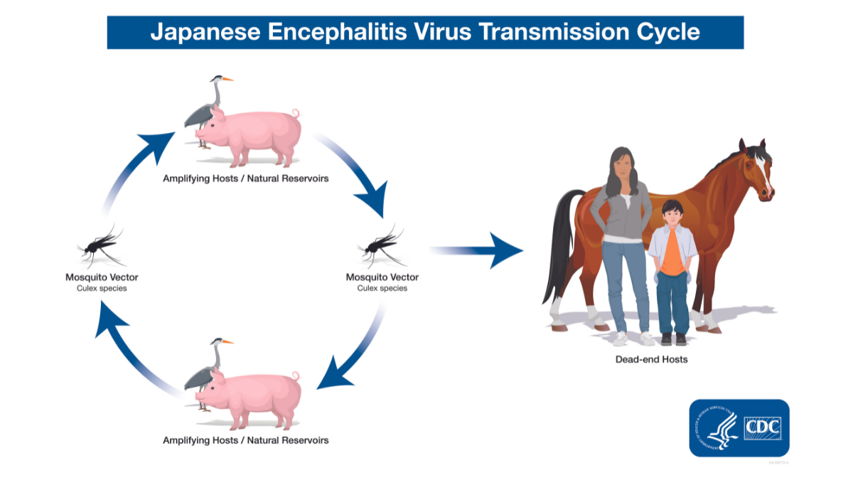
- Transmission strictly follows an enzootic cycle involving mosquito vectors and vertebrate hosts.
| Transmission Component | Specific Details |
|---|---|
| Primary Vector | Transmitted primarily by Culex tritaeniorhynchus mosquitoes, which breed prolifically in stagnant water bodies like rice paddies. |
| Natural Reservoirs | Ardeid birds, such as herons and egrets, naturally maintain the virus in the environment. |
| Amplifying Hosts | Pigs serve as the most critical amplifying hosts, developing high-titer viremia sufficient to infect feeding mosquitoes. |
| Dead-End Hosts | Humans and horses represent incidental, dead-end hosts because they fail to develop sufficient viremia to infect mosquitoes. |
Pathophysiology
- Following an infected mosquito bite, the virus undergoes local replication and enters regional lymph nodes, triggering a transient viremia.
- Neuroinvasion occurs across the blood-brain barrier via passive transport, infected leukocyte transit, or olfactory neuronal entry.
- The virus causes direct neuronal destruction and provokes an intense inflammatory response featuring perivascular lymphocytic cuffing and microglial nodules.
- Neuropathology characteristically localizes to the thalamus, basal ganglia, and brainstem.
Clinical Manifestations
- The standard incubation period ranges from 4 to 14 days.
- While over 99% of infections remain completely asymptomatic, clinical illness progresses through three distinct symptomatic stages.
| Clinical Stage | Duration And Key Features |
|---|---|
| Prodromal Stage | ➤ Lasts 2 to 3 days; presents with abrupt high-grade fever, chills, severe headache, anorexia, and vomiting. |
| Acute Encephalitic Stage | ➤ Lasts 3 to 4 days; features progressive clouding of consciousness, delirium, and coma. ➤ Generalized tonic-clonic seizures manifest frequently in pediatric patients. ➤ Extrapyramidal signs represent a distinctive hallmark, presenting as mask-like facies, cogwheel rigidity, dystonia, and choreoathetoid movements. |
| Convalescent Stage | ➤ Lasts weeks to months; fever subsides, but persistent neurological sequelae remain highly common. |
Diagnostic Evaluation
| Diagnostic Modality | Characteristic Findings |
|---|---|
| Serology | Cerebrospinal fluid or serum IgM Capture ELISA represents the gold standard diagnostic method, successfully detecting antibodies within 7 days of symptom onset. |
| Neuroimaging | Magnetic Resonance Imaging demonstrates characteristic bilateral hyperintensities on T2 and FLAIR sequences prominently involving the thalamus and basal ganglia. |
| Cerebrospinal Fluid | Analysis typically reveals mild to moderate lymphocytic pleocytosis, mildly elevated protein, and strictly normal glucose levels. |
Management Protocol
- No specific antiviral therapy currently exists; clinical management remains strictly supportive.
- Airway patency must be rigorously secured, occasionally requiring intubation and mechanical ventilation for comatose patients.
- Raised intracranial pressure mandates head elevation to 30 degrees and the administration of hyperosmolar therapy like Mannitol.
- Meticulous fluid monitoring is essential to detect and properly manage concurrent Syndrome of Inappropriate Antidiuretic Hormone secretion.
- Seizures demand aggressive control utilizing intravenous Benzodiazepines, immediately followed by Phenytoin administration.
Prognosis And Prevention
- The disease carries a high mortality rate ranging from 20% to 30%, occurring primarily due to raised intracranial pressure or acute respiratory failure.
- Permanent neurological or psychiatric sequelae, including severe motor deficits and intellectual impairment, afflict 30% to 50% of disease survivors.
- Universal vaccination constitutes the single most effective preventive strategy.
- The live attenuated SA 14-14-2 vaccine is routinely administered in two doses at 9 months and 16 to 24 months of age under the Universal Immunization Program in highly endemic districts.
- Environmental control necessitates the strict segregation of pigs from human habitats and rigorous vector control targeting rice paddies.